

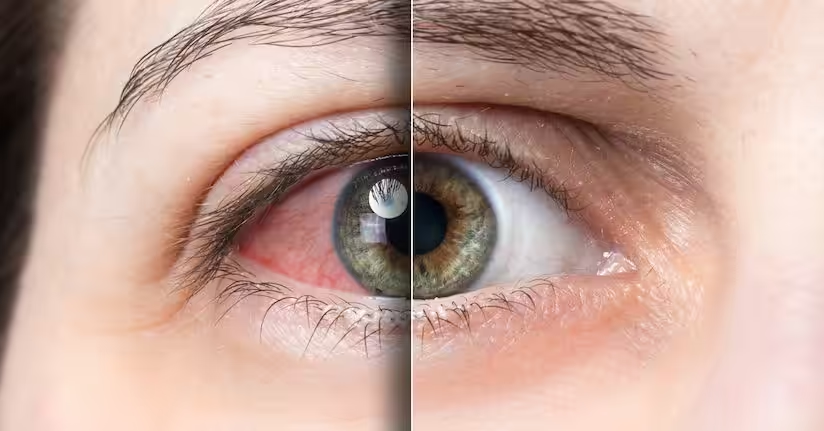
Dry eye is a chronic symptomatic condition which, if not addressed promptly and appropriately, can lead to ocular surface disease. It is estimated that approximately 100,000,000 people suffer from it worldwide. Often, it is underdiagnosed and remains untreated, while in other cases it is underestimated and treated only symptomatically with tear substitutes and/or corticosteroids, depending on the case.
The disease is multifactorial, and the loss of integrity of the tear film initially affects visual function to a limited extent, while in severe cases it may even lead to vision loss.
Revised Definition:
Dry eye is a multifactorial disease of the ocular surface characterized by the loss of homeostasis of the tear film and accompanied by ocular symptoms. Etiological factors include tear film instability and hyperosmolarity, inflammation, ocular surface damage, as well as various neurosensory abnormalities.
The new definition allows the inclusion of subclinical conditions that involve ocular surface changes without the presence of symptoms.
The complex and sensitive system of ocular surface homeostasis is affected by various environmental and habitual factors, hormonal changes, age, multiple local ocular and systemic diseases, as well as the use of both topical and systemic medications.
Symptoms vary in intensity and type and are not easily distinguishable between the different categories of dry eye, making diagnosis challenging.
Chronically affected patients are not easily diagnosed and often use various formulations without significant or long-term effect.
Today, a decisive step in management is recognizing the specific dysfunctions of the components responsible for maintaining ocular surface homeostasis.
These components include:
a) Eyelids – their position, functionality, blink quality and frequency, and the glands producing the lipid layer of the tears
b) Conjunctiva – with goblet cells that produce mucin
c) Limbal region (sclero-corneal border) – responsible for epithelial renewal of the cornea with its specific microenvironment
d) Cornea – with its rich neural network
e) Lacrimal apparatus – including the secretory glands of the aqueous layer and the lacrimal drainage system
f) Sensory and motor innervation of the above structures
Based on the cause and resulting dysfunction, a stepwise approach to management is determined, aiming ultimately to improve the patient’s quality of life.
The approach to correct diagnosis includes a complete history (habits and occupation), a questionnaire, a stepwise and organized examination of the structures maintaining ocular surface health, a summary of results, and management based on the cause, resulting dysfunction, and severity of findings.
It is important for ophthalmologists to acquire both theoretical knowledge and practical skills because there is no single diagnostic test to confirm the presence of dry eye and ocular surface disease (OSD).
Combining various diagnostic tests increases the accuracy of diagnosis, leading to better management.
Understanding the pathophysiological mechanisms, knowledge of different examination methods, and integrated presentation of results will lead to diagnosis and initial proper management.
The following analysis outlines the steps for diagnosis and management as a guide to improving the approach and treatment of these patients.
INSTRUCTIONS:
Diagnosis and Management of Ocular Surface Disorders Defined as Dry Eye and Blepharitis
STEPWISE DIAGNOSIS:
aims to
Identify patients suspected of developing ocular surface disorders (OSD)
Safely diagnose patients with OSD
Determine the causes of OSD
Manage dysfunctions and improve patients’ quality of life
Educate and inform both patients and other healthcare providers involved in ocular surface dysfunctions to achieve comprehensive management
1. When surface damage is primary and tear production is very low, diagnosis is made using the Schirmer test and the Oxford scale is used to grade severity. In this case, aqueous tear deficiency from the lacrimal gland is investigated for Sjögren syndrome and non-Sjögren syndrome.
2. Tear film instability disorders, along with findings from Meibomian glands and eyelashes, indicate increased evaporation.
3. Exogenous factors such as contact lens use, eye drops, ocular surface disease, and environmental factors are investigated regarding the cause and type of disorder.
DIAGNOSIS/ASSESSMENT
History
Patient demographics
History of symptom onset, duration, exacerbations and remissions, previous treatments, and symptom progression over time
Investigation of habits, occupation, environmental factors, and their effect on symptoms
Complete ophthalmic history including surgeries, use of eye drops, and contact lenses
Complete medical history and history of systemic medications
Questionnaire: OSDI (Ocular Surface Disease Index) – the most widely used. Helps with initial symptom assessment and is used during management to monitor treatment response.
Evaluation of Symptoms/Findings
The primary diagnostic symptom of dry eye is intermittent blurred vision.
There may be a discrepancy between symptoms and findings, either in early stages (many symptoms with few findings) or in advanced stages (many findings with few symptoms).
Ophthalmic examination is targeted to diagnose tear and ocular surface disorders.
Tear film and ocular surface evaluation via slit-lamp examination
Tear quantity assessment: Schirmer test (<5 mm reliable) or phenol red thread test
Fluorescein Assessment:
Staining with Lissamine Green/Rose Bengal
Fluorescein/Rose Bengal/Lissamine Green characteristics
Stains healthy cells | No | Yes | No |
Stains dead/degenerating cells | No | Yes | Yes |
Staining blocked by mucin layer | No | Yes | No |
Toxicity | No | Yes | Yes |
Real diffusion rate through collagen | Very fast | Very slow | Fast |
Coloring through cell junction breakdown
Tear film protection insufficient, dead/degenerating cells, cell junction breakdown
Oxford scale: Corneal and conjunctival staining grading
Tear meniscus height assessment (TMH)
Fluorescein clearance test: Tear drainage assessment
Tear film stability: TBUT (tear breakup time)
Tear film dynamics (TF-dynamics): thinning/movement time
Conjunctival folds/looseness (LIPCOF – CCH)
Tarsal and bulbar conjunctiva assessment and redness grading, including limbal evaluation
Other diagnostic tests:
Tear osmolarity measurement
Tear interferometry
MMP-9 (metalloproteinase 9) – ocular surface inflammation marker
Inflammatory Tear Marker: Measurement of MMP-9 in tears is an objective indicator of inflammation on the ocular surface. The test has 92% specificity and 87% sensitivity for diagnosing the presence of dry eye. In a positive test, the patient is likely to respond to anti-inflammatory therapy. It is also used to monitor the effectiveness of treatment.
Impression cytology
Mucin assessment (tear ferning test)
Tear protein analysis
Eyelid and Meibomian gland examination
Eyelid and Lid Margin Assessment:
Observation of eyelid position, shape, skin texture, facial appearance, and potential rosacea (cheek telangiectasia, rhinophyma, etc.)
Assessment of eyelid functionality: mobility, closure pattern
Horizontal lid laxity evaluation (medial-lateral canthal ligaments), lower lid retractors, floppy eyelid check
Blink assessment: frequency (increased e.g., dry eye, decreased e.g., Parkinson’s), quality; incomplete blinking reduces Meibomian gland expression and lipid layer distribution, increasing tear evaporation
Eyelid Margin Examination via Slit-Lamp:
Evaluation of eyelash quality, quantity, and position; check for Demodex at base of lashes
Lid margin shape and position relative to globe; normal: smooth trapezoid in contact with globe; puncta internally positioned without eversion on closure
Inflammation signs: hyperemia, telangiectasia
Meibomian gland orifices: visible, smooth, without secretion, or with abnormal secretion/atrophy
Meibomian Gland Evaluation:
Meiboscopy: assesses gland presence, dilation, or atrophy
Meibography (Keratograph 5M): records gland morphology, classifies pre-existing damage
Function evaluation: MGEvaluator or manual expression to assess quality/quantity of meibum: clear, cloudy, granular/paste-like
Pressure applied: mild, moderate, or firm; MGEvaluator ensures consistent pressure
Count of functional glands per lower lid segment
Diagnostic and staging devices for follow-up and treatment efficacy
MANAGEMENT / TREATMENT
Eyelid hygiene: foams, gels, or impregnated pads; selection based on treatment goals
Topical therapy:
Tear substitutes (artificial tears), tailored to type, severity, and patient response
Suppression of ocular surface inflammation:
Cyclosporine 0.5–1% eye drops, 2–4 times daily
Autologous serum 20%
Mild corticosteroids (fluorometholone or hydrocortisone), varying dose and duration, careful for side effects
Antimicrobials (tobramycin, gentamicin, chloramphenicol) for active blepharitis
Improving Meibomian Gland Secretion Quality:
Azithromycin
Tetracycline/doxycycline systemic
Omega-3 supplementation
TTO (tea tree oil) or impregnated wipes for coexisting Demodex
Warm compresses and gland expression
Blinking exercises
Surgical interventions:
Conjunctival laxity correction
Punctal eversion
Entropion/ectropion/floppy eyelid correction
Tarsorrhaphy for exposure
Corrective eyelid/conjunctiva/lacrimal surgeries
Amniotic membrane or limbal stem cell transplant for ocular surface disease
Punctal plugs
Alternative therapies:
Hydrophilic contact lenses as a bandage
Specialized glasses to create a high-humidity ocular chamber
Environmental and systemic factors:
Identify and regulate environmental triggers
Address systemic issues (thyroid, hormone replacement, autoimmune diseases)
Patient education:
Chronicity explanation and treatment rationale encourage compliance
Treatment selection is based on findings and patient cooperation
Realistic patient expectations
Ongoing discussion with patient regarding symptom evolution
Follow-ups: Regular follow-up visits aim to assess the effect of treatment on both symptoms and ocular surface findings. Any lesions and changes resulting from the therapy are re-evaluated, and appropriate questions are used to investigate the treatment’s impact on the patient’s quality of life. The intervals between follow-up visits are adjusted according to the patient’s response to the proposed therapy. Follow-ups may be frequent initially (e.g., every few days) and gradually become less frequent (once or twice a year) after the condition stabilizes.
Dalianis, A. Karidis, V. Liolios
Edited by: Ch. Terzidou