
The macula is the central part of the retina, located at the posterior pole of the eye. It is responsible for central vision and for the ability to perceive the fine details that make up everything we see.
Macular degeneration is a condition that essentially causes alterations to this part of the retina — the macula — resulting in blurring of the patient’s central vision as well as difficulty in perceiving visual stimuli.
Symptoms often vary among patients. In the early stages of macular degeneration, there may be no symptoms at all. However, during a comprehensive ophthalmologic examination, the condition can be detected even in patients who have shown no prior indications.
The main symptoms are:
Blurred vision
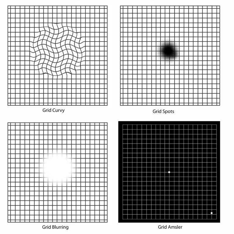
Central scotoma (loss of central vision)
Difficulty reading and driving
Difficulty recognizing faces
Decreased visual acuity
Distorted vision (metamorphopsia)
Need for brighter lighting
Finally, in advanced stages of macular degeneration, vision loss may occur.
Age-related macular degeneration primarily affects individuals over 60 years old. However, a significant factor in the development of the disease is genetic predisposition, which can also lead to symptoms appearing at a younger age. In addition to age and family history, a modern unhealthy lifestyle (Western-type diets) plays an important role in the onset of the condition. Specifically, smoking, diet, and prolonged exposure to ultraviolet radiation from sunlight appear to affect the macula and constitute risk factors for this disease.
The two main forms of age-related macular degeneration (AMD) are:
Dry type (non-exudative or atrophic form):
This is the most common and generally the least dangerous form of AMD, as its progression is slow, resulting in a gradual loss of vision. It is primarily caused by thinning, atrophy, and aging of the macula. Dry-type macular degeneration is characterized by yellowish and white deposits of lipids and proteins (drusen) that accumulate between the retina and the choroid. There is no targeted treatment for this form of AMD, apart from certain specialized dietary supplements. In advanced stages, the patient may experience significant reduction in visual acuity and may require visual aids, such as magnifying lenses, to assist with daily activities.
Wet type (exudative form):
This form of age-related macular degeneration affects approximately 10–15% of patients. In wet AMD, pathological new blood vessels develop beneath the retina due to a factor called VEGF (vascular endothelial growth factor). These vessels are responsible for causing edema and hemorrhages, which lead to vision loss. Wet AMD can be treated or managed with intravitreal injections of anti-VEGF agents that block VEGF and limit the formation of new vessels and edema. This approach stabilizes visual acuity in patients and, in many cases, can even improve it.
Dry-type AMD can occasionally progress to the wet type.
For the diagnosis of age-related macular degeneration (AMD), the following examinations are essential:
Fundoscopy with pupil dilation: Examining the fundus of the eye can reveal the presence of lesions and drusen (deposits), which are characteristic features of AMD.
Measurement of visual acuity.
Amsler test: This test is printed on a card featuring a grid of horizontal and vertical lines with a central dot. Patients with dry or wet AMD may perceive gaps or wavy lines when viewing the grid.
Optical coherence tomography (OCT): This imaging technique highlights retinal lesions in the macular region.
Digital angiography (Angio-OCT) or angiography with intravenous indocyanine or fluorescein: These procedures provide detailed information about the veins and arteries inside the eye.