


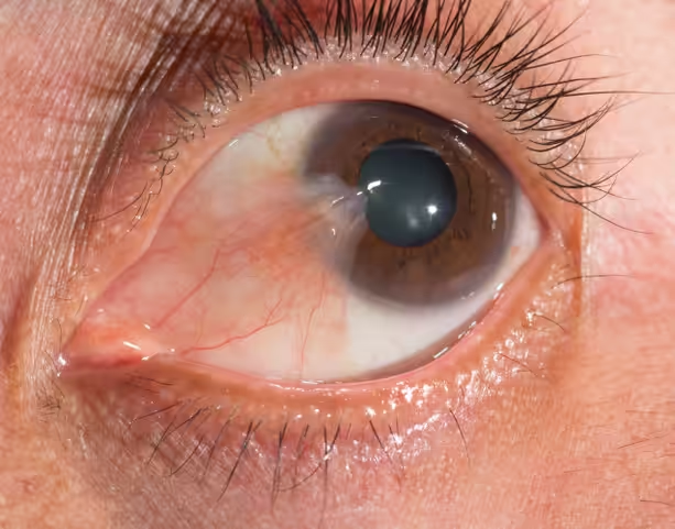
Pterygium is an eye condition in which a fibrovascular tissue grows over the eye, usually extending toward the center of the cornea. This tissue often originates from the inner corner of the eye (nasal side) and, because it covers the sclera (the white part of the eye), it can interfere with vision and often causes refractive errors, such as increased astigmatism. This degenerative change of the conjunctiva occurs gradually, and its size varies in each case.
The main causes of pterygium include:
Symptoms vary depending on the size and extent of the fibrovascular growth. They may include:
The management and treatment of pterygium is carried out either locally with the use of eye drops and ophthalmic ointments, or surgically in cases where the visual axis is threatened with a weakening of the patient’s vision. Of course, surgery is also performed for cosmetic reasons if the extension of the fibrovascular tissue is significant. During the surgical procedure, local anesthesia is applied.
The surgical technique that will ensure permanent treatment in most cases is determined individually.
If the pterygium is small but tends to extend toward the visual axis, a simple excision and suturing of the conjunctiva (the transparent tissue covering the white of the eye) can be performed using a Z-plasty technique.
In large pterygia, a longer surgical procedure may be performed, which, to prevent recurrence, will require a conjunctival autograft and adhesion with surgical glue. The autograft is taken from the superior conjunctiva (under the upper eyelid).