


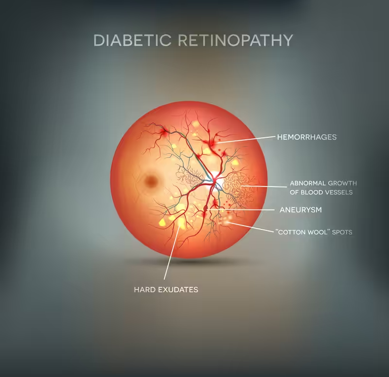
Diabetic retinopathy is an eye disease in which lesions (damage) occur in the retina of diabetic patients. This condition mainly affects individuals who have had high blood sugar levels for a prolonged period or those who have been living with diabetes for many years. Essentially, the small blood vessels of the retina are affected, leading to their dysfunction, the formation of new abnormal vessels (neovascularization), and tissue necrosis (ischemia) in the areas they supply with blood.
There are two types of diabetic retinopathy: non-proliferative (NPDR) and proliferative (PDR).
Non-proliferative diabetic retinopathy is primarily an early stage of the disease. At this stage, hemorrhages, microaneurysms, exudates, and retinal edema can be observed in the fundus of the eye. When any of these occur in the central retina, that is, in the macula, the patient develops macular edema. This edema significantly affects the patient’s visual acuity and is also an indicator of disease severity.
However, in non-proliferative diabetic retinopathy, patients may have no visual loss or only mild visual impairment (when macular edema is not present).
Proliferative diabetic retinopathy is the most severe form of the disease. At this stage, new blood vessels develop on the surface of the retina (neovascularization). This occurs due to poor blood circulation in the eye (ischemia) caused by the obstruction of the retina’s normal blood vessels. Prolonged ischemia triggers the production of a protein called VEGF (vascular endothelial growth factor), which in turn promotes the formation of new vessels. These new vessels develop on the retinal surface and are abnormal. Because of their extremely thin walls, they are fragile and prone to leakage. Their rupture causes bleeding either into the vitreous or at the level of the retina. If these vessels grow in the anterior part of the eye, they may block the normal drainage of aqueous humor (the angle), leading to neovascular glaucoma. In advanced stages, scarring or even tractional retinal detachment may occur, both of which can result in severe vision loss.
Blurred vision
Floaters or flashes of light
Double vision
Fluctuating vision
It is also possible that, in the early stages of diabetic retinopathy, none of the above symptoms may yet be present.
Regulating and closely monitoring blood sugar levels, combined with systematic eye examinations and early diagnosis by an ophthalmologist, are key to preventing and avoiding diabetic retinopathy.
Based on the diagnosis and the patient’s condition, the most appropriate course of treatment and management is determined.
The main treatment methods include:
Anti-angiogenic (anti-VEGF) agents:
Injectable therapy that inhibits the action of the protein known as VEGF, which is responsible for the growth of fragile, abnormal blood vessels (neovascularization) on the surface of the retina. Anti-VEGF agents also improve macular edema that may have developed.
Intravitreal corticosteroid injections (in various forms)
Photocoagulation – application of specialized laser beams to the retina:
These laser beams destroy and cauterize the pathological new vessels, helping to reduce fluid leakage.
Surgical treatment – Vitrectomy:
Surgery is indicated in cases with complications such as retinal detachment or vitreous hemorrhage.
Finally, the retinal vessels are the only microcirculatory vessels that can be directly observed through a simple examination (fundoscopy). Similar vessels exist in the kidneys and other organs. Therefore, potential retinopathy may also indicate more serious systemic complications of diabetes affecting other organs.