


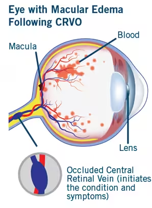
Veins are the blood vessels that carry blood away from the retina toward the central circulation. In some cases, these veins can become blocked. The occlusion may affect a branch of the retinal vein or its central trunk. When these vessels are obstructed, a cascade of events occurs that can impair retinal function in the affected area.
Risk Factors
As with many vascular conditions, risk factors for retinal vein occlusion include: High blood pressure, advanced age, hyperlipidemia, diabetes mellitus, elevated intraocular pressure, and smoking.
Studies have also shown that contraceptive medications can increase the risk of retinal vein occlusion, along with other predisposing conditions such as chronic kidney failure, dehydration, and hyperviscosity syndromes.
The primary mechanism of retinal vein occlusion is pressure exerted by the wall of a neighboring artery on the adjacent vein at the point where they cross. This typically occurs due to arterial wall hardening.
The interruption of blood flow caused by this compression can lead to the formation of a clot.
What are the symptoms of retinal vein occlusion?
The primary symptom is vision loss, which can be partial or complete depending on the location of the occlusion.
Central vision impairment is caused by macular edema, the main complication of the condition.
Patients with central retinal vein occlusion are at increased risk of developing neovascularization (formation of abnormal new blood vessels) in the following months. This can lead to neovascular glaucoma, which causes painful elevation of intraocular pressure and further vision deterioration. These complications arise from extensive retinal ischemia due to reduced blood flow.
Diagnosis is made through a clinical eye examination, specifically fundoscopy. Optical Coherence Tomography (OCT), digital retinal angiography, or traditional fluorescein angiography, help provide the most accurate and comprehensive diagnostic approach.
What is the treatment for retinal vein occlusion?
Management of retinal vein occlusion requires attention to the patient’s overall health, as it is a vascular condition. Addressing any associated systemic diseases—such as diabetes, hypertension, hyperlipidemia, or metabolic syndrome—is crucial.
Intravitreal Anti-VEGF Injections
These medications treat macular edema caused by the occlusion. The number of injections varies by patient, as each case is unique.
Intravitreal Steroid Implants
Steroid implants are highly effective for managing macular edema secondary to retinal vein occlusion. Potential drawbacks include increased intraocular pressure in some patients and a risk of cataract formation.
Laser Therapy
In cases of neovascularization (identified via fluorescein angiography), retinal photocoagulation laser is the treatment of choice. The laser targets areas of the retina, reducing oxygen demand and causing regression of abnormal new vessels.
General Guidelines
For all patients with central or branch retinal vein occlusion blood pressure, lipid profile, and blood sugar levels ought to be monitored. A cardiological evaluation is necessary, as are some specialized hematologic tests which are likely to identify underlying causes of the disease