


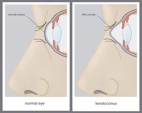
Keratoconus is an eye condition in which the normal structure of the cornea progressively thins, forming a cone-shaped cornea. This alters the quality of vision and gradually reduces visual acuity. The condition usually affects both eyes.
This disorder most often appears in the mid-second decade of life. Its initial onset may go unnoticed by the patient or the ophthalmologist. The first sign is the continual, frequent change in the patient’s eyeglass prescription, primarily due to increasing astigmatism. In advanced stages, vision can be significantly reduced.
Keratoconus is a rare corneal disorder. It occurs in approximately 1 in 2,000 people, with no geographic predilection. The exact causes of keratoconus remain unknown. However, research indicates that a family history is the most significant risk factor (heredity).
Other studies suggest a correlation between the progression of the disease and conditions that cause itching (allergies or atopic disorders), particularly during childhood.
Diagnosis is made by the clinical ophthalmologist through corneal topography examination. In this test, a computer is used to analyze the structure of the cornea and generate a map of its anterior and posterior surfaces.
The treatment of keratoconus has two main objectives: