


Glaucoma is a serious, multifactorial disease. It is the most common cause of irreversible vision loss (4.4 million – 12.3% of the blind population worldwide). It affects 2% of white adults over the age of 40 and occurs four times more frequently in African Americans. Due to the aging of the global population, the prevalence of glaucoma is continuously increasing. It is worth noting that most patients do not know they have glaucoma.
Causes – risk factors
Increased intraocular pressure (IOP) is closely associated with glaucoma. The general public tends to think that a patient with high eye pressure necessarily has the disease. However, this is not exactly the case. A healthy eye can sometimes present with high intraocular pressure (ocular hypertension), while an eye with completely normal pressure can have advanced glaucoma. Nevertheless, intraocular pressure remains the most significant measurable risk factor.
In general, glaucoma risk factors are divided into two major categories: modifiable and non-modifiable. Increased IOP and corticosteroid use belong to the first group, while age, family history (DNA), myopia, thin corneal thickness, and history of trauma comprise the non-modifiable risk factors for glaucoma.
What causes ocular hypertension?
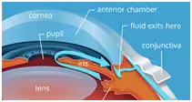
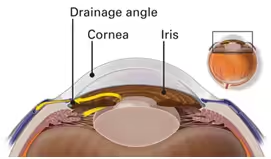
The aqueous humor is the fluid that fills the anterior part of the eye. It is produced by the ciliary body, located behind the iris. The aqueous humor passes through the pupil and circulates in the anterior chambers of the eye. Its drainage occurs through the anterior chamber angle, formed by the cornea (the transparent window of the eye) and the iris. When intraocular pressure (IOP) is normal, there is a balance between the fluid being drained and the fluid being produced. When drainage is affected or obstructed, intraocular pressure naturally increases.
Glaucoma – symptoms
Many patients believe that a sensation of heaviness or retrobulbar pain is due to increased eye pressure. The most common form of glaucoma, chronic simple glaucoma, gives absolutely no symptoms, which is why it is called “the silent thief of vision.”
Vision loss begins at the periphery, losing some details of the image without the patient noticing. In the final stages, vision becomes tunnel-like or there is total blindness.
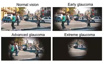
The only type of glaucoma that occurs suddenly with severe pain, nausea, blurred vision, and eye redness is acute angle-closure glaucoma, which is much rarer than chronic open-angle glaucoma.
Types of glaucoma
Chronic simple glaucoma or open-angle glaucoma
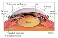
In this type, the drainage of aqueous humor is open, but there is increased resistance to outflow through the trabecular meshwork. It is the most common and insidious form of glaucoma, as it is completely asymptomatic.
Angle-closure glaucoma (acute glaucoma)
Occurs when the angle between the cornea and iris is completely closed. Patients with high hyperopia and older individuals are more likely to develop it. It is usually accompanied by nausea, vomiting, or severe headache. It is an ophthalmic emergency and must be treated as soon as possible.
Sub-acute angle-closure glaucoma
In this type, the patient suffers from angle-closure attacks, which may often go unnoticed. The iris mechanically blocks the natural drainage of the anterior chamber, and the resulting waves of increased pressure gradually damage the optic nerve. Quite often, these patients experience severe pain, especially in dim light, or see halos around bright sources.

Normal-tension glaucoma
In some patients, the disease occurs even when intraocular pressure is within normal or even low levels. Women, elderly patients, those with nocturnal systemic hypotension (especially those on antihypertensive therapy), and patients with sleep apnea syndrome are at higher risk. The presence of vascular phenomena, such as Raynaud’s syndrome, has also been implicated as a risk factor.
Pigmentary glaucoma
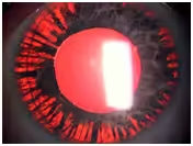
Pigment dispersion syndrome is a condition in which pigment cells from the back of the iris travel with the flow of aqueous humor through the pupil and block the anterior chamber angle, the main drainage pathway of the eye. This results in increased intraocular pressure and, over time, the development of glaucomatous damage (pigmentary glaucoma).
In this type of glaucoma, intraocular pressure can fluctuate significantly, reaching very high levels (40 mmHg). Nevertheless, not all patients with pigment dispersion syndrome will develop glaucoma. About 10% of patients develop glaucoma within 10 years, and 15% within 15 years.
Neonatal or congenital glaucoma (childhood glaucoma)
Glaucoma in children is very rare. However, it can appear immediately after birth or during the first months or years of life (congenital glaucoma). It affects about 1 in 10,000 newborns.
It is usually due to a malformation or dysfunction of the anterior chamber angle, where aqueous humor drains. This occurs due to improper development during the fetal period.
The most important sign for early diagnosis is buphthalmos. The term buphthalmos indicates enlargement of the infant’s or child’s eye due to ocular hypertension. In children with very high pressure, corneal edema may also be present, causing it to appear cloudy. Additionally, these young patients may have tearing and photophobia.
Glaucoma treatments
One in two glaucoma patients in Greece does not know they have it!
Thus, the most important treatment for glaucoma is prevention. Ophthalmic examinations should be performed every two years after age 40, and if risk factors exist, annual or more frequent check-ups are recommended.
The main modifiable risk factor for glaucoma is intraocular pressure; therefore, the primary goal of treatment is its reduction. Initial therapy can be performed with eye drops or laser.
Eye drops
Eye drops can regulate pressure by either reducing aqueous humor production or increasing its outflow. Proper technique and frequency of instillation are very important for correct patient management. Elderly patients with significant musculoskeletal or rheumatologic conditions may have difficulty properly instilling the drops, making the treatment ineffective. Poor compliance, when the patient does not understand the risks of uncontrolled ocular hypertension and does not use their medication regularly, can also render treatment ineffective.
Laser
Glaucoma lasers have a unique role in treatment. They can be used as a first-line therapy if the patient prefers to avoid eye drops or has difficulty administering them. Laser treatment has two primary goals:
- reducing aqueous humor production
- increasing its outflow
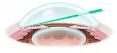
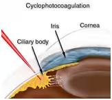
The leading-edge lasers used in glaucoma treatment are SLT (Selective Laser Trabeculoplasty), sub-Cyclo Laser, and endo-cyclo-photocoagulation laser.
When all the above are ineffective, surgical treatment of glaucoma is considered, either via conventional glaucoma surgery or implantation of a drainage valve.
MIGS
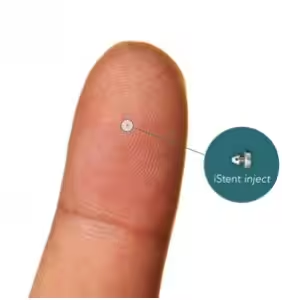
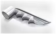
New surgical treatments introduced in recent years have significantly improved both surgical outcomes and patient recovery time. These are MIGS (Minimal Invasive Glaucoma Surgery). These treatments are highly effective for moderate glaucoma or ocular hypertension cases where large reductions in IOP are not necessary.
Surgeries
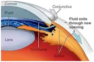
Trabeculectomy – Deep Sclerotomy – Valves
For glaucoma cases that do not respond to the above treatments and require greater pressure regulation, the progression can be halted through surgical techniques such as trabeculectomy, deep sclerotomy, or valve implantation. These techniques divert aqueous humor from the anterior chamber to the suprachoroidal space, lowering intraocular pressure and thereby halting glaucoma progression.
These are glaucoma-specific surgeries that require special expertise, have a significant recovery period, and can provide important solutions in disease management.
Myths and truths
Diet and glaucoma
There is no correlation between diet and glaucoma. One type of glaucoma, neovascular glaucoma, primarily caused by diabetes, may require dietary regulation—not to manage glaucoma itself, but to control the underlying disease, which is diabetes.
Cannabis – is it a good treatment for glaucoma?
An increasing number of countries promote medical cannabis for chronic conditions. Cannabis can temporarily lower intraocular pressure, but the required dose would be extremely high, potentially threatening the patient’s physical and mental health. Therefore, NO, cannabis should not be used to treat glaucoma.
Glaucoma specialization
One of the most important causes of vision loss is inadequate diagnosis and insufficient treatment.
In the past, glaucoma assessment relied on a simple measurement of eye pressure. Those days are gone.
All patients should be systematically examined by an ophthalmologist specializing in glaucoma.
Necessary ophthalmologic examinations for glaucoma include:
- Tonometry and tonometric curve
- Gonioscopy
- Measurement of central corneal thickness
- Evaluation of the optic nerve and nerve fibers
- Visual field testing
Finally, patient education regarding disease management, adherence to treatment, and periodic monitoring of the optic nerve are what ensure safe stabilization of the condition.
“A well-informed patient does not ask about their eye pressure, but about the state of their optic nerve.”
Eye Center